Original songlei Shanghai children’s medical center ophthalmology

Why do you want to do fundus screening?
Premature babies are prone to retinopathy of prematurity (ROP), which will develop into serious blinding eye diseases if not treated in time. Premature infants leave their mothers when their organs are not fully developed. At this time, the development of retinal blood vessels is not mature. After birth, due to the change of blood oxygen concentration, abnormal vascular proliferation is prone to occur. Abnormal vascular rupture will lead to retinal hemorrhage, fibrous proliferation and contraction, retinal detachment, etc., and even blindness and eyeball atrophy in severe cases. ROP must be found and treated early. If treated in time, the effect is quite good.

Which children need screening and when?
For premature infants and low birth weight infants with birth weight less than 2000g, the first screening should be conducted at 4-6 weeks after birth or at 32 weeks after gestational age correction. The screening scope of premature infants with serious diseases can be appropriately expanded. The frequency of follow-up is generally determined according to the fundus lesions: once every 2 weeks without ROP, once every 1~2 weeks when ROP is diagnosed, and the interval of ROP follow-up before the threshold should be less than 1 week. Complete vascularization of retina, static lesion, scarring, or having received treatment are the termination time of screening and follow-up for retinopathy of prematurity.

What do you think of ROP inspection results?
Diagnostic criteria divide the retina into three areas:
Zone ⅰ: the central area of the retina in the circle with the optic disc as the center and the radius of twice the distance from the optic disc to the fovea maculata.
Zone II: that annular area of retina with the optic disc as the cent and the distance from the optic disc to the serrated edge of nose as the radiu.
Zone ⅲ: the temporal semilunar retinal region.
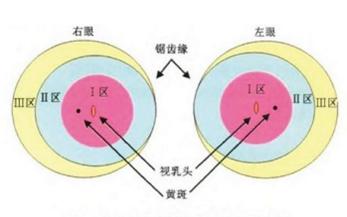
The zoning indicates the location of the lesion,
The lesion in area ⅰ is the most serious, and the lesion in area ⅲ is mild.
According to the severity of ROP lesions, it is divided into five stages, and the general lesions develop from stage 1 to stage 5 (as shown below). When the disease reaches stage 4, there is little hope of recovery through surgery.
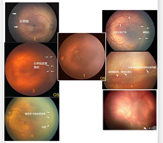
There is a dividing line between the vascular area and the avascular area around the temporal side of ROP in stage 1.
Stage 3 ROP PLUS(+) not only has crista-like protuberance, but also has additional pathological changes: tortuous dilatation of blood vessels, which is more serious than that in Figure 3.
The boundary of ROP in Phase 2 is widened and uplifted.
Normal fundus
Partial retinal detachment caused by contraction and traction of ROP fibroproliferative membrane in stage 4
Stage 3 ROP cristae, accompanied by hyperplasia and hemorrhage.
Stage 5 ROP total retinal detachment
According to the above classification criteria, the baby with ROP will be recorded in the medical record: ROP in several districts and periods, PLUS(+or-). (Note: PLUS is an additional lesion, showing tortuous dilation of retinal vessels, which is a manifestation of rapid progress of ROP)

Screening process
During hospitalization, the baby’s fundus examination will be arranged by the ward; You can go to the outpatient clinic for examination after discharge.
Outpatient service process:
1. After the attending doctor determines the need for fundus screening, he will prescribe medicine for examination.
2. After the parents pay for the medicine, they start to give the baby some mydriatic liquid medicine. Generally, it takes one hour from the beginning of the medicine to the examination of mydriasis.
3. 1~2 drops of surface anesthetic water will be given before the examination, so that the baby can be examined in a painless state.
4. During the examination, the baby was lying on the bed, and the doctor opened the eyelids with a special eyelid opener for the baby. The retina was examined with binocular indirect ophthalmoscope and wide-angle children’s fundus photography system, and recorded according to the lesion area and lesion degree.
5. The doctor will inform whether treatment is needed and/or the time for the next review according to the examination results.
6. After the baby receives the examination, it needs to use anti-inflammatory eye drops for 3 days.

friendly reminder
Please try to arrive at 13: 00 ~ 14: 00 because you need some eye drops and wait for mydriasis before the examination, otherwise your waiting time will be affected.
In order to avoid aspiration pneumonia or even suffocation caused by excessive milk during examination, parents are requested to estimate the time and stop feeding after starting to order medicine.
You need to hold your baby’s head tightly and fix it during the examination. Please ask parents to understand and cooperate. Generally, the examination time only takes 5~10 minutes. Don’t interrupt or give up the examination because of the crying of children and the distress of adults. If the blindness caused by this happens, you will regret it for life.
ROP emphasizes early detection and early treatment. Early intervention and treatment can achieve normal children’s vision. If found late, it may lead to irreversible blindness. Since 2009, our team has carried out fundus screening for premature babies, with more than 2,500 screenings every year, which has accumulated rich experience, and we will give timely and standardized treatment according to the screening results.
The ophthalmology department of Children’s Center has opened a specialized clinic for newborns and children with basic diseases. Please rest assured that our team will take care of your baby’s bright eyes with you.
Read the original text